Dr Hany Elmadbouh, Consultant Musclo-skeletal and Image-guided Intervention explain the value of using imaging to guide inection.
Clinicians from different disciplines perform musculoskeletal injections to treat pain.
Pain is one of the most frequent causes of reductions in productivity as a result of sick leave and decreases in workers’ ability to carry out their jobs. Each year around a fifth of the population consult their GP about a musculoskeletal condition, which accounts for £5 billion of NHS spend and is the leading cause of working days lost. Arthritis Research UK estimates its costs the economy £20 billion every year. Treating all types of back pain alone costs the NHS more than £1 billion per year. These figures highlight the economic importance and strong social impact of the growing use of low-cost interventions capable of producing appreciable, long-lasting pain relief.
Most joint and tissue injections can be performed via the visualization and palpation of anatomical landmarks to guide appropriate placement. However, blind injections carry the risk of incidental needling or drug delivery to the adjacent non-target structures, which may include blood vessels, peripheral nerves, muscles, ligaments, intra-tendinous tissue and sub-cutaneous fat, cannot be completely avoided.
Many injections are given using only anatomical landmarking and although there has been an increase in the use of ultrasound guided injections by predominantly Rheumatologists for whom injection therapy is the most common therapeutic intervention, it is the Interventional Radiologists who use image guidance without exception in their work. Perhaps the chief reason for the increased use of image guidance has been the improved technology. Better visualization of anatomy yields greater confidence to undertake procedures that were previously thought to be dangerous or impossible. Also, there has been significant progress in materials and in biology that lead to more effective interventions. Improvements in the speed of image acquisition and in 3-D technology are changing the way that procedures are performed and making new procedures possible.
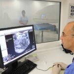
Ultrasound has become an increasingly valuable tool for guiding musculoskeletal interventions due to improvements in probes and image- processing software. Ultrasound offers the added advantage of portability, low cost, availability and use on non ionizing radiation.
Image guided joints and soft tissue diagnostic and/or therapeutic injections can also be performed under Fluoroscopy, CT and more recently MRI. In general, the majority of injections are performed using ultrasound or fluoroscopies which are more efficient than CT. The exception is spinal injections which are more readily done under CT. The choice of the imaging modality may be dependent on availability, the expertise of the clinician performing the injection, the characteristics of the patient and the clinical problem.
In general, ultrasound is used for soft tissue lesions, fluoroscopy for superficial bone lesions and joints injections, and CT with or without CT fluoroscopy for spine injections.
Image guidance offers a number of advantages. It helps to confirm the indication/contraindication of the procedure. With image-guidance, it is possible to plan an accurate and safe access route. Image guided procedures are less painful than blind injection. The accurate needle and treatment positioning, may lead to better clinical outcome.
At Avicenna clinic, we have a range of specialist consultants, operating theatre for surgical procedures and superior in-house imaging facilities –including state-of-the-art MRI, ultrasound and X-ray scanning equipment. We can assess and diagnose all cases of acute and chronic pain quickly and deliver comprehensive treatment plans tailored to you.
To book a consultation or for more information on treating your pain and explore the other available services to help you, contact Avicenna Clinic on 0330 202 0597.